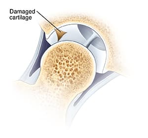
When the firm, smooth tissue that covers the ball and socket of the hip is damaged, it can cause painful catching in the joint. This tissue is called cartilage. Injury to cartilage is called chondral damage. Arthroscopy is a surgery method that uses only small incisions and small tools. It can be done to fix chondral damage.
In the operating room
Just before surgery, you may be asked several times which hip is to be treated. This is a standard safety measure in the operating room. You will likely receive medicine (anesthesia) to make you sleep.
During the procedure
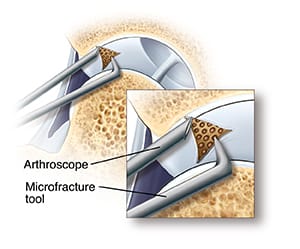
After you receive anesthesia, your leg is gently pulled to widen the hip joint. Next, the surgeon makes a few small incisions called portals. Through these portals, they insert surgical tools, including the arthroscope. The arthroscope sends images of the joint to a screen. These images allow the surgeon to look inside the joint. The joint is filled with sterile fluid to help the surgeon see more clearly.
Repairing chondral damage
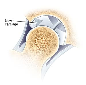
If the damaged cartilage is loose, it is removed. If the cartilage is missing, the exposed bone may be shaved to smooth it. Or small holes may be placed in the bone (microfracture). This allows new cartilage to form. Once the surgeon finishes, the portals are closed and bandaged. Then you are taken to the recovery room.

Orthopedics CTA
Find a DoctorRelated Articles
Microdiskectomy
During a microdiscectomy, some disk is removed. In most cases, bone must first be removed to expose the damaged disk. The part of the disk outer wall and soft center that presses on the nerve can then be removed. There is usually enough disk remaining to cushion the vertebrae.
Kneecap Surgery: Removing Damaged Cartilage
Surgery may be used when pain severely limits your activities. Or it may be done when a rehab program or other nonsurgical treatments just are not helping enough. Some procedures may be done using arthroscopy. This method uses tiny incisions and special instruments to look and work inside the knee joint. Other procedures need open surgery.
Understanding Clavicle Fracture Open Reduction and Internal Fixation (ORIF)
Open reduction and internal fixation (ORIF) is a type of treatment to fix a broken bone. It puts the pieces of a broken bone back together so they can heal.
Foot Surgery: Curled Fifth Toe
A curled fifth toe is most often inherited. When the fifth toe curls inward, it moves under the next toe. Then the nail of the curled toe starts to face outward. As a result, you may bear weight on the side of your toe instead of the bottom. This can cause corns and painful nails. There are many nonsurgical treatments available. But if these are not effective, surgery is a choice.